Heel neuroma: the enigma of recalcitrant heel pain and an innovative approach highlighting sixty surgical cases and a review of two hundred and fifty-seven symptomatic but non-surgical cases
Ira D. Shandles, James Pruchniewski and Katy L. Reynolds
The authors report resolution of recalcitrant heel pain by describing new insights into innervation of the heel. They elucidate biomechanical mechanisms responsible for entrapment of this innervation and explain the diagnostic and therapeutic techniques necessary to manage this etiology, including an innovative therapeutic approach. A one-to-ten year follow-up of the course of 317 heel pain syndrome patients was conducted. The first 216 cases were treated by basic technology providing a cure, with 161 patients responding to a conservative approach and 55 requiring surgery. The final 101 cases received Diapulse® combined with their conservative care and 96 responded while only five required surgery. A 95% success rate of cure was achieved using the new technology without surgery. The sixty surgical cases are proposed as confirmation of the diagnosis because all are accompanied by irrefutable pathological reports. All surgical cases preoperatively were in turn subjected to the same diagnostic and therapeutic protocols as the non-surgical.
Introduction
Heel neuroma and its correction is offered as the solution to the heel pain problem frequently seen in podiatric medicine (Beito et al. 1991). The differential diagnoses offered for this symptom have included calcaneal spur, bursitis, plantar fascitis, calcaneal stress fracture, osteomyelitis, rheumatic and seronegative arthropathies, as well as cutaneous manifestations, fat pad atrophy, neoplasms, vascular insufficiency or vascular congestion, and L-5 to S-1 radiculopathy (Leis et al. 1986, Jennings et al. 1988, Kulund 1988). A biomechanical fault is found to exist in almost all heel pain patients and its control is an important element in their management (Root et al. 1977).
When conservative therapy fails, patients may undergo heel spur resection with plantar fasciotomy (Murphy & Baxter 1985, Jennings et al. 1988, White 1994). This procedure is frequently unsuccessful, the length of recovery long, and morbidity high (Przylucki & Jones 1981, Savastono & Jones 1981, Murphy & Baxter 1985, Baxter et al. 1989) as authors recognized the need for a better understanding of the etiology for recalcitrant heel pain in order to explain the unacceptable rate of disappointment.
Heel neuroma and its correction is offered as the solution.
Researchers and surgeons worldwide have successfully, over the past fourteen years, dissected and elucidated the exact branch of the tibial nerve responsible for the onset of a heel neuroma (Przylucki & Jones 1981, Murphy & Baxter 1985, Savastono 1985, Baxter et al. 1989, Weil 1994). The questions became:
- From what etiology can this neuroma’s appearance be attributed?
- By what easily reproducible means can its presence be demonstrated?
- How should it be systematically treated?
Understanding the anatomy
The consensus of independent researcher surgeons has been that the unique branch of the tibial nerve involved in the process of heel neuroma development is some part of the first branch (FB) of the lateral plantar nerve (Przylucki & Jones 1981, Murphy & Baxter 1985, Baxter et al. 1989, Weil 1994).
The reluctance to accept nerve entrapment as a source of heel pain may stem from a misconception regarding the course of the FB of the lateral plantar nerve, placing it well distal to the usual area of heel pain near the medial portion of the calcaneal tuberosity (Baxter et al. 1989). Recent studies show that the FB bifurcates from the lateral plantar nerve more proximally than was formerly believed, and as it passes across the heel it innervates the periosteum overlying the medial tuberosity of the calcaneus (Weil 1994) (Figs 1a & b).
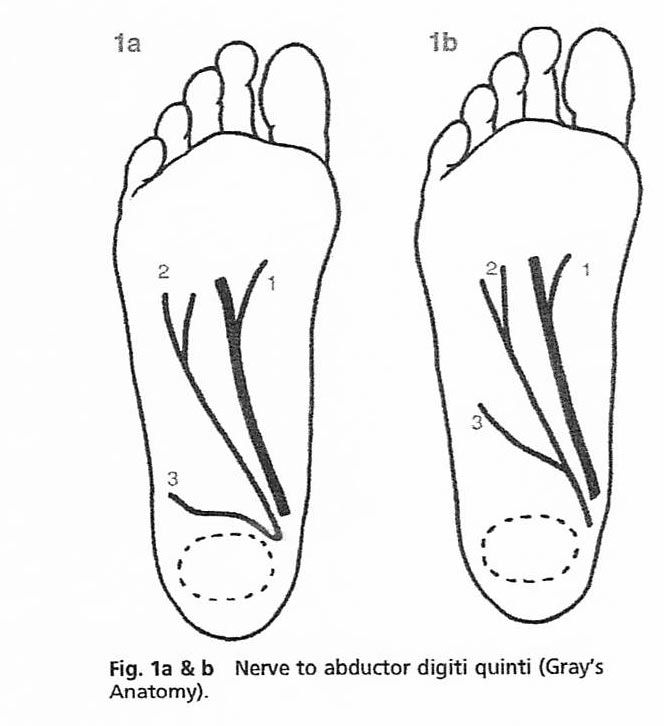
The FB is comprised of three separate divisions:
- The first, to the medial portion of the calcaneal tuberosity — medial and lateral perichondrium included.
- The second, supplying the flexor digitorum brevis.
- The third, supplying the abductor digiti quinti brevis (Baxter et al. 1989, Weil 1994) (Fig. 2).
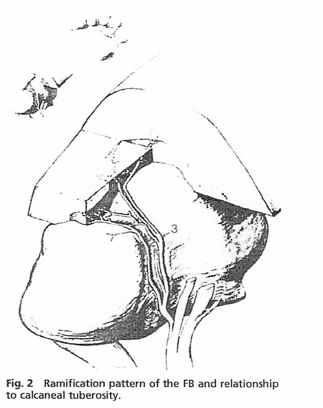
Fortunately, the neuroma’s body appears to be principally isolated to the division innervating the medial portion of the tuberosity and associated perichondrium, which is the first branch of the first branch of the lateral plantar nerve, a purely sensory branch, superior to, but contiguous with the region above the developing inferior calcaneal spur. It is important to envision that when this calcaneal branch leaves the first branch anatomically, it runs due plantarward to a branch of the laciniate ligament on the medioplantar aspect of the heel before sharply turning due lateralward into the medial portion of the tuberosity of the calcaneus, a superficial position, before reaching its final deep destination. Therefore, understanding the anatomy of the entire FB explains why the pain tends to occur about the heel plantarly, medially and laterally, and is sometimes associated with radiations both dorsally and proximally, and even along the lateral margins of the foot. The FB, by its very anatomy, solves the riddle of the source of these varied but associated focal complaints.
Biomechanics
Biomechanics explains the etiology of the tumour once the existence of the precise branch of the peripheral nerve tissue is accepted. Pronation secondary to compensated forefoot or rearfoot varus, flexible forefoot valgus or rearfoot valgus, and compensated equinus can all contribute to heel pain (Root et al. 1977, Davis et al.). It has been suggested that plantar fascia can create pressure against nerves resulting in ‘pronation neuritis’ (Kulund 1988). Hyperpronation will also create compression where the FB passes the medial portion of the calcaneal tuberosity.
A heel spur or inflammation at the origin of the plantar fascia could produce enough swelling to compress the FB against the long plantar ligament and calcaneus during the stance phase of gait (Baxter et al. 1989). Recurrent irritation can lead to chronic neuritis and ultimately hypertrophy of the nerve sheath fibres (Beito et al. 1991).
The challenge for the clinician is to reproduce the neuroma-type pain near the medial junction of the heel in order to make the differential diagnosis from true fasciitis or ‘bursitis’. The phenomenon of simple plantar fasciitis should be reproducible easily upon forced passive dorsiflexion of the foot at the level of the metatarsal heads upon the leg, without palpation of the fascial band (Kulund 1988). If this manoeuvre fails to reproduce the plantar heel pain of which the patient is so aware, fasciitis is unlikely.
Materials and methods
Patient population
Patients averaged 45 years of age, with lesion distribution 39.1% in males and 60.9% in females. Fifteen pediatric cases were identified clinically in this study. It may also be noteworthy that the average adult patient was found to be very heavy, averaging 206 lbs.
All patients had an initial complete podiatric history and physical examination concurrent with the diagnostic examination which led to localization of the pathology to the involved tissue.
Examination
- The blunt end of a definitive object with a diameter of about 1/4 inch, such as the eraser end of a pencil, is placed against the medioplantar aspect of the heel with moderate pressure.
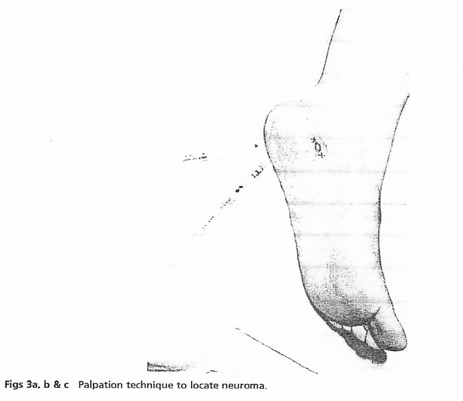
- The blunt probe is pressed in a discrete manner against successive sites along this area marking an ‘X’ at points at which pain (not pressure-sense) is elicited.
- At one point the patient will indicate that the pain is most severe. Mark that point with an ‘O’ (Figs 3a, b & c).
- From the point ‘O’, palpate superiorly slightly and inferiorly slightly to determine that this is truly the most sensitive spot.
- Upon surgical dissection, the point ‘O’ has proven to be in its central aspect in direct proximity to the tumour 100% of the time, and from this point injection therapy can be initiated.
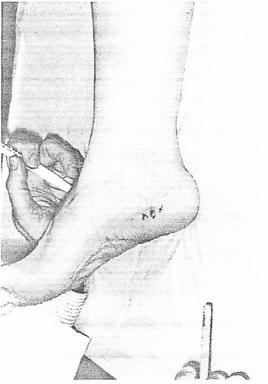
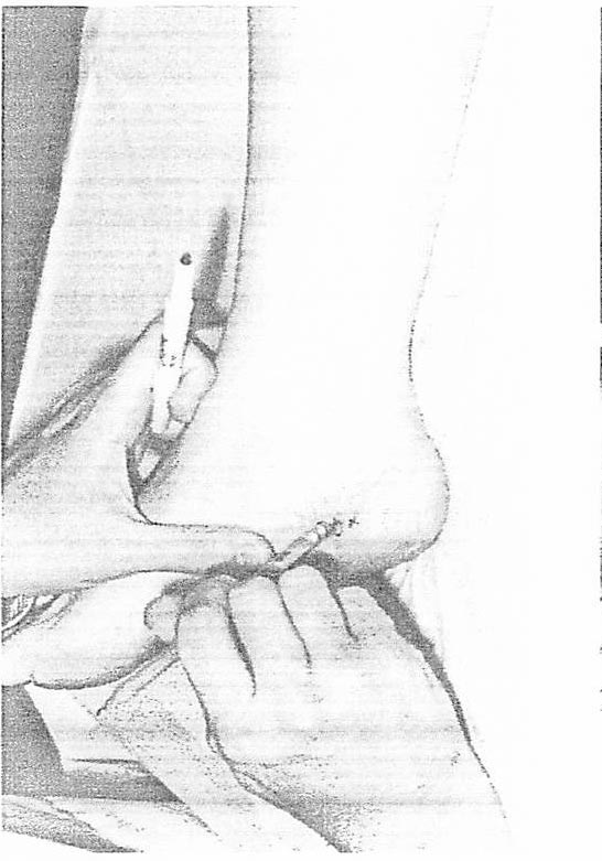
Figs 3a, b & c. Palpation technique to locate neuroma.
The differential diagnosis should include a full array of metabolic or other orthopedic disorders which could produce heel pain — especially if a bilateral case (Leis et al. 1986) is being considered.
Diagnostic Materials
The 1990s have brought two additional non-invasive and definitive diagnostic modalities which can be used to test for the presence or absence of this lesion, providing greater certainty and illustration: neurometer and lumbar thermography.
- The neurometer is a neuroselective diagnostic stimulator which enables physicians to obtain a quantitative measure of nerve integrity from three different nerve groups (A, B, and C fibres). One electrode is placed over the site marked ‘O’ and the other dorsally directed towards the forefoot tibial nerve at the level of the tarsal canal. An abnormal reading should be consistently achieved.
- Lumbar thermography by a certified technician using strict Wexler protocol and one of the accepted standard models of equipment, the tumour is readily visible. Its nature is inflammatory; it will usually glow at the highest ends of the color scale in a discrete heel location (Fig. 4). There are very few exceptions; this study consistently provided visual confirmation of the presence of the lesion.
Once the tumour has been differentiated, a decision regarding conservative treatment can be made.
Treatment technology: Non-surgical
In our study, one choice was to follow the approach of injection therapy which had been found to be effective in the preponderance of forefoot neuromas: 0.2 cc of Decadron Acetate 8 mg/cc, 1 cc of vitamin B12, and finally 0.3 cc of lidocaine 2% plain were combined. The injection was delivered most comfortably using a 27 gauge needle after numbing the skin with a skin refrigerant such as Ethyl Chloride* or Fluroethyl*. The mixture was shaken and infused slowly. Injections should be spaced at least one week apart. As the tumourous tissue is quite dense, it can be felt on injection and should be discovered superficially 1/8 to 1/4 inch below the skin surface.
During the last five years of the study, injections were followed immediately by a Diapulse® (Great Neck NY) treatment to enhance penetration and absorption. The injection therapy combined with Diapulse therapy is responsible for the statistical significance and overall success of the procedure without surgery. The first five years of this study were performed by administering injections alone without this technology and success was achieved in only 75% of the patients. During the last five years, when a single Diapulse treatment was administered to the affected heel(s) as well as to the epigastrium, the success rate increased to better than 95%.
Diapulse produces pulsed high frequency high peak power electromagnetic energy, with a maximum output of 975 watts of instantaneous power in 65 microsecond athermic bursts (Diapulse® Corporation of America). It has been well researched; its efficacy for the reduction of edema and pain with no contraindications or side effects has been reported (Kaplan & Weinstock 1968, Erman 1970, Richmond 1970, Hersh 1972, Barclay et al. 1982, Pennington et al. 1993). The therapy is non-thermal, non-invasive, painless, safe and the energy penetrates surgical dressings and plaster casts. Treating the epigastrium improves peripheral blood flow. Additional accelerated wound healing has been documented.
With strict adherence to the aforementioned differential diagnostic scheme and the injection and treatment procedure outlined, a consistent success rate can be anticipated in the majority of patients with this conservative approach.
In the rare case in which the injection therapy fails, the use of orthoses as a second hedge against surgery is usually not recommended. Normally, the orthosis should be looked upon as a mandatory follow-up to a successful course of injection therapy, since the likelihood of it providing relief in the face of a chronically inflamed, and probably fibrosed, nerve tissue, is nil.
In pediatric cases where injection therapy was undesirable, physical therapy using ice packs thirty minutes daily, or, during acute phases, moist heat along with physical massage and Diapulse treatment three times weekly, was substituted. This was usually combined with NSAIDs for three or four weeks.
Treatment technology: Surgical
The importance of listening to each patient at the time of surgery must be stressed emphatically. In each case, even under the influence of pre-operative sedation, patients were quite responsive to the palpatory exam and gave information regarding location of the tumour. In each case, it was found under the mark made on the skin prior to the procedure.
In this study of 317 cases, a total of 60 required surgery, 55 pre-Diapulse and five post-Diapulse. All surgical cases of neuroma were subsequently verified histologically with changes virtually identical with the classic Morton’s neuroma. All surgical specimens were subjected to routine pathological analysis by Board Certified Pathologists.
The surgery is technically simple, because the tumour is very superficial: a slightly curvilinear incision was made parallel to the plantar aspect of the foot. The incision was then deepened via sharp and blunt means down to the hypodermis. The tumour should be identified in the centre of the field as described by the patient when the diagnostic tool has been used.
The tumour can be differentiated from the surrounding normal fat by the arrangement of fibrous tissue overlying it. The fibrous bundle referred to appears as a nummular portion of laciniate ligament overlying an even deeper, firmer, fatty tissue which encompasses the neurofibril elements. The fibrous patch is then grasped with a small Allis forceps, circumscribed, and dissected via CO₂ laser set at eight to ten watts pulsed at half second intervals, to include the deeper, firm, related tissues, both laterally, superiorly and finally deeply down to the tuberosity of the calcaneus (Figs 5a & b).
The final mass ranged in size from 1 × 2.5 cm to 1.5 × 3.5 cm. As dissection progressed, the healthy, resilient, neighbouring fatty tissue was clearly distinguishable. Once the tumourous mass was excised, the surrounding tissue was easy to reapproximate and the dead space adequately filled, using skin closure only because of the voluminous supple fat in the area. Masses larger than those described might require the placement of a suction drainage system. The skin was closed with 2-0 or 3-0 nylon or prolene suture, applied in horizontal mattress and simple interrupted style (Figs 6a & b). A large fluff dressing was secured to the wound and surrounded by wrapping a 4″–6″ Ace* bandage from toes to knee.
All surgical patients were treated on an out-patient basis and received the non-invasive Diapulse treatment at each postoperative visit in the last five years of the study. Treatment was administered two to three times per week, settings 600 pulsations/6 penetration for 10–15 minutes over the area of involvement, and 400/4 for 10–15 minutes over the epigastrium. This method accelerates healing without contraindications or side effects (Erdman 1960, Hedenius et al. 1966). Patients were kept at limited ambulation for the first 48 h; permitted to ambulate in a post-operative shoe, and all sutures were removed on the tenth post-operative day. By the end of the third week, most patients were able to return to normal shoe gear with comfort, preferably with an orthosis in place.
Root Functional Orthoses were used post-operatively. The Root Laboratory (Auburn CA) provided all corroborative forefoot measurements based on neutral position casts of the respective patients.
Within the first 48 h after surgery, patients reported nearly complete disappearance of the pain they felt pre-operatively, noting that any pain felt at this point was clearly related to the surgical procedure. Diapulse, in the last five years of the study, reduced the pain, thus diminishing the requirement for most analgesics. All patients were cured although those treated with Diapulse did not require surgery as frequently: 20% fewer patients required surgery.
Results
It is important to understand that all patients were cured in this study, with over 95% to the injection/Diapulse and the balance with surgery/Diapulse during the last five years. The one-to-ten year follow-up has shown a complete cure of the syndrome in the 257 cases with the use of the injection therapy (and Diapulse treatments in the last five years of the study). In those cases, managed conservatively, between one and five injections were administered — although up to eleven injections were required in one particularly resistant case.
For a comprehensive review of the data arising from this study, see Table 1. The attached Flow Sheet (Table 2) demonstrates the appropriate diagnostic and treatment criteria for heel pain based on the current research.
The surgical cases, all of which had failed to respond completely to the aforementioned injection therapy, demonstrated 100% relief of symptoms within a few days to thirteen weeks of surgery combined with Diapulse therapy to enhance healing and pain relief. Six of the cases in the entire group of 317 were bilateral (Tables 3 & 4).
In this study, biomechanical evaluation of all unilateral cases demonstrated an unequal distribution between compensated forefoot varus and flexible forefoot valgus foot types (69.5% varus vs 37.9% valgus). Regardless of etiology, subtalar joint pronation in excess of normal pronation appeared in all cases to be the common denominator. It is interesting to note that the neuroma syndrome demonstrated itself on the foot with greater pronatory capacity only 40.9% of the time.
Although this apparent contradiction cannot be explained conclusively, it seems logical to suggest that when pronation exceeds a certain point, the position of the FB of the lateral plantar nerve, with reference to the plantar topography of the calcaneus and surrounding anatomy of the rear foot, assumes a less irritating course on the more pronated side.
The charts and graphs indicate the statistical significance of the study. The results demonstrate the beneficial physiological and biophysical significance of our method.
Discussion
In the differential diagnosis of heel pain, heel neuroma is all too commonly overlooked, however, it should be considered as the major contributor. Heel neuroma is readily identifiable from other causes of heel pain. Once the program is established, 75% of patients (or as many as 95% when Diapulse is part of the treatment program) should respond to appropriate conservative measures as outlined in this report. Surgical excision should be reserved for those resistant cases that do not respond to conservative care. The problem should prove to resolve uniformly, particularly with the adjunctive use of Diapulse to accelerate healing thus diminishing scar tissue formation.
This research covered a ten-year follow-up of data collected and collated from medical records, by the authors. The problem of heel pain is well studied and diagnosed: heel neuroma and its correction is offered as the solution. Careful diagnosis should lead to a successful treatment programme and offer patients long-term relief. The above described method avoids complications, is an office and out-patient procedure, and provides a minimum of discomfort for the patient with an expected outcome.
In the opinion of the authors, this work conclusively proves that the use of a custom orthotic device, following a successful course of injection therapy or surgery, can prevent a recurrence of the original heel pain problem, even after ten years of weight-bearing activity. The adjunctive use of Diapulse significantly and statistically enhances the outcome.
The preceding report has portrayed the heel neuroma syndrome in all its aspects as a tangible, treatable, and curable entity.