
Historical clinical paper · Sixth International Congress on Burns · August 31, 1982 · San Francisco, California
Agrippa Ionescu, M.D.; Doina Ionescu, M.D.; Stefan Milinescu, M.D.; biologist Elvira Talnar; and Silvia Vieru, M.D. Clinic of Plastic Reconstructive Surgery, Bucharest, Romania; Biochemical Laboratory of the Post Graduate Medical School, Fundeni Hospital, Bucharest.
Introduction
Starting in 1974, the authors report using pulsed high-frequency electromagnetic energy, described as Diapulse therapy, in patients with oedema, pain, burns, traumatic lesions, surgery, inflammatory disease, necrotic lesions, and radionecrosis. They state that early clinical results led them to investigate biochemical indicators of tissue viability.
The paper focuses on epidermal proteins and enzymes. It cites earlier work on enzymes in normal skin and describes the roles of oxidative enzymes, glycolysis, carbohydrate metabolism, and transaminases in epidermal tissue. The authors chose local enzymatic activity as an indicator of tissue viability and compared proteins plus lactate dehydrogenase (LDH), glutamic-oxalacetate transaminase (GOT), glutamic-pyruvic transaminase (GPT), and alkaline phosphatase in normal and burned tissue, before and after the reported therapy.
Materials and methods
The paper describes a device operating at a radio frequency of 27.12 MHz, with pulsed output from 80 to 600 times per second. The authors report treatment once daily over each burned area for 10 minutes at a setting of 600 pulses per second and six peak-power steps.
The study included 20 patients with burns admitted as emergencies. Three 100–300 mg skin samples were taken from each patient: one from the burned area before treatment, one from a nearby unburned area as a control, and one from the burned area 24 hours after the reported therapy. Samples were washed in saline, homogenized, centrifuged, filtered, and analyzed for proteins, LDH, GOT, GPT, and alkaline phosphatase by spectrophotometry.
Results
The concentrations of proteins and enzymes in healthy tissue, and burn tissue, treated with Diapulse therapy is reproduced on Table 1.
Complete case data
(Table 1)
Open a section to review the full source dataset for the corresponding tissue condition.
Normal tissue — complete 20-case data
| Case | Proteins | GOT | GPT | Alk. Ph. | LDH |
|---|---|---|---|---|---|
| 1 | 1.138 | 43.94 | 30.04 | 1.75 | 163.4 |
| 2 | 1.187 | 106 | 18 | 1 | 917 |
| 3 | 0.644 | 161.49 | 38.82 | 15.52 | 875.7 |
| 4 | 0.608 | 273 | 49.34 | 21.38 | 1097.3 |
| 5 | 0.568 | 237.67 | 61.62 | 12.32 | 1378.5 |
| 6 | 1.350 | 28.88 | 9.28 | 3.70 | 822.2 |
| 7 | 2.906 | 98.76 | 11.69 | 3.09 | 465.5 |
| 8 | 1.666 | 90.03 | 9.6 | 6 | 567.82 |
| 9 | 3.310 | 80.96 | 11.48 | 4.53 | 568.8 |
| 10 | 1.046 | 37.28 | 32.94 | 7.65 | 672.8 |
| 11 | 1.230 | 19.07 | 14.63 | 11.63 | 478.86 |
| 12 | 1.564 | 142.58 | 7.67 | 3.19 | 802.43 |
| 13 | 0.533 | 118.19 | 11.25 | 15.62 | 819.88 |
| 14 | 1.928 | 119.8 | 6.22 | 13.20 | 948.65 |
| 15 | 1.222 | 35.18 | 9.82 | 13.91 | 652.20 |
| 16 | 1.804 | 118.07 | 10.53 | 11.64 | 648 |
| 17 | 1.359 | 95.65 | 16.92 | 14.71 | 710.67 |
| 18 | 1.224 | 100.65 | 15.15 | 4.33 | 809.52 |
| 19 | 1.190 | 143.70 | 24.37 | 19.32 | 857.98 |
| 20 | 1.137 | 114.33 | 21.10 | 10.55 | 872.3 |
Burned tissue — complete 20-case data
| Case | Proteins | GOT | GPT | Alk. Ph. | LDH |
|---|---|---|---|---|---|
| 1 | 0.756 | 178.57 | 18.75 | 6.61 | 121.3 |
| 2 | 1.009 | 43.94 | 10.54 | 0.87 | 78.20 |
| 3 | 1.138 | 30.75 | 14.94 | 1.75 | 96.66 |
| 4 | 0.382 | 196.3 | 28.79 | 20.94 | 643.97 |
| 5 | 0.657 | 101.97 | 22.83 | 10.65 | 663.62 |
| 6 | 1.724 | 33.06 | 6.05 | 1.74 | 311.4 |
| 7 | 2.261 | 7.96 | 3.98 | 1.32 | 122.5 |
| 8 | 1.702 | 42.30 | 5.28 | 4.11 | 192.71 |
| 9 | 2.817 | 18.46 | 3.55 | 1.06 | 135.25 |
| 10 | 0.737 | 59.70 | 23.06 | 5.42 | 324.50 |
| 11 | 0.624 | 16.02 | 20.83 | 4.80 | 172.11 |
| 12 | 2.022 | 79.13 | 7.91 | 10.38 | 649.35 |
| 13 | 0.408 | 51.47 | 9.80 | 7.35 | 276.96 |
| 14 | 1.621 | 49.21 | 4.57 | 6.48 | 505.91 |
| 15 | 0.768 | 22.03 | 14.32 | 10.41 | 281.25 |
| 16 | 1.155 | 112.76 | 12.99 | 16.24 | 622.73 |
| 17 | 1.226 | 79.12 | 26.91 | 16.31 | 657.42 |
| 18 | 1.090 | 76.14 | 13.76 | 2.75 | 575.23 |
| 19 | 1.119 | 53.62 | 20.55 | 17.87 | 412.86 |
| 20 | 0.799 | 90.11 | 17.52 | 7.51 | 504.25 |
Diapulse-treated tissue — complete 20-case data
| Case | Proteins | GOT | GPT | Alk. Ph. | LDH |
|---|---|---|---|---|---|
| 1 | 1.333 | 23.25 | 16.50 | 6.00 | 348.8 |
| 2 | 0.810 | 72.55 | 25.49 | 1.96 | 58.82 |
| 3 | 1.333 | 20.25 | 9 | 6.75 | 183.79 |
| 4 | 0.244 | 49.18 | 4.09 | 16.39 | 459 |
| 5 | 0.537 | 199.25 | 22.35 | 18.62 | 877.09 |
| 6 | 2.097 | 30.52 | 6.67 | 3.81 | 300.9 |
| 7 | 2.475 | 15.75 | 3.63 | 2.02 | 545.4 |
| 8 | 2.981 | 40.25 | 6.37 | 2.01 | 471.98 |
| 9 | 1.111 | 12.60 | 7.20 | 7.20 | 206.1 |
| 10 | 0.913 | 19.71 | 24.24 | 25.19 | 479.8 |
| 11 | 1.164 | 24.91 | 17.73 | 10.31 | 376.28 |
| 12 | 2.399 | 71.69 | 4.16 | 9.58 | 622.76 |
| 13 | 1.719 | 69.22 | 5.81 | 11.63 | 399.65 |
| 14 | 1.928 | 47.71 | 11.92 | 6.22 | 502.44 |
| 15 | 2.222 | 24.6 | 4.50 | 20.25 | 456.79 |
| 16 | 1.826 | 93.09 | 10.40 | 20.81 | 558.59 |
| 17 | 1.564 | 54.98 | 7.03 | 31.96 | 698.8 |
| 18 | 0.826 | 78.70 | 75.06 | 14.52 | 707.02 |
| 19 | 1.235 | 97.16 | 22.67 | 18.62 | 652.63 |
| 20 | 1.186 | 101.18 | 20.24 | 12.65 | 877.1 |
Average values of concentration of proteins and enzymes referred to Proteins (Micro/mgP) in health and burned tissue before and after Diapulse therapy.
(table 2)
| Measure | Normal tissue | Burned tissue | Diapulse-treated tissue |
|---|---|---|---|
| Proteins | 1.38 | 1.20 | 1.49 |
| GOT | 108.26 | 67.13 | 57.33 |
| GPT | 20.52 | 14.34 | 15.25 |
| Alkaline phosphatase | 9.75 | 7.73 | 12.32 |
| LDH | 756.47 | 367.41 | 489.20 |
We note in the case of proteins, GPT, alkaline phosphatase and LDH, a decrease in burned tissue versus healthy tissue, i.e., for proteins, the decrease is from 1.38 Iu/mgP in healthy tissue to 1.20 Iu/mgP in epidermal burned tissue; — 13%.
Source: Ionescu et al., Sixth International Congress on Burns, 1982.
—GPT decreases from 20.52 Iu/mgP in healthy tissue to 14.34 Iu/mgP in burned tissue; — 30%.
—Alkaline phosphatase decreases from the average value of 9.75 Iu/mgP in the control tissue to 7.74 Iu/mgP in the burned tissue; — 21%.
—LDH decreases from 756.47 Iu/mgP to 367.41 Iu/mgP; — 51%.
After Diapulse therapy we note a return to normal; even greater values for proteins, GPT, alkaline phosphatase and LDH.
Versus burned tissue, the proteins increased 19%, the average value in burned tissue treated by Diapulse therapy (1.49 Iu/mgP) versus healthy tissue (1.38 Iu/mgP). GPT increased 6% in treated tissue versus burned tissue.
Alkaline phosphatase increased 37% in treated tissue versus burned, the average value after treatment (12.32 Iu/mgP), higher than the average value in healthy tissue (9.75 Iu/mgP).
All the same, LDH increased 25% in treated tissue versus burned, the average value after Diapulse therapy (489.20 Iu/mgP) being lesser than the average value in healthy tissue (756.47 Iu/mgP).
Differing from proteins and the other three enzymes, GOT decreases 38% in burned tissue versus the healthy one and continues to decrease after Diapulse therapy in the burned tissue, 57%.
Percent variations of the average concentration of protein GOT, GPT, alkaline phosphatase and LDH in burned tissue versus normal tissue and in the Diapulse treated versus burned tissue.
(Table 3)
| Measure | Normal / burned | Diapulse-treated / burned |
|---|---|---|
| Proteins | −13% | +19% |
| GOT | −38% | −57% |
| GPT | −30% | +6% |
| Alkaline phosphatase | −21% | +37% |
| LDH | −51% | +25% |
DISCUSSION
In the case of burned tissue compared to healthy tissue, proteins decreased by 13%, GOT decreased by 38%, GPT decreased by 30%, alkaline phosphatase decreased by 21% and LDH decreased by 51%.
In the case of burned tissue treated with Diapulse compared to burned tissue untreated, proteins increased by 19%, GPT increased by 6%, alkaline phosphatase increased by 37% and LDH increased by 25%. Differing from proteins and the other 3 enzymes, GOT continued to decrease from -38% to -57%.
The ratios of the concentrations of proteins and enzymes in treated tissue versus those in burned tissue are noted in Table 4.
TABLE 4
| No. cases | Proteins | GOT | GPT | Alkaline phosphatase | LDH |
|---|---|---|---|---|---|
| 1 | 1.76 | 0.13 | 0.88 | 0.90 | 2.87 |
| 2 | 0.80 | 1.65 | 2.42 | 2.25 | 0.75 |
| 3 | 1.17 | 0.66 | 0.60 | 3.85 | 1.90 |
| 4 | 0.64 | 0.25 | 0.14 | 0.78 | 0.71 |
| 5 | 0.82 | 1.95 | 0.98 | 1.75 | 1.32 |
| 6 | 1.21 | 0.92 | 0.10 | 2.19 | 0.96 |
| 7 | 1.09 | 1.98 | 0.91 | 1.53 | 4.45 |
| 8 | 1.75 | 0.95 | 1.20 | 0.49 | 2.45 |
| 9 | 0.39 | 0.68 | 2.03 | 6.79 | 1.52 |
| 10 | 1.24 | 0.33 | 1.05 | 4.64 | 1.48 |
| 11 | 1.86 | 1.55 | 0.85 | 2.15 | 2.19 |
| 12 | 1.18 | 0.90 | 0.53 | 0.92 | 0.96 |
| 13 | 4.21 | 1.34 | 0.59 | 1.58 | 1.44 |
| 14 | 1.19 | 0.97 | 2.60 | 0.96 | 0.99 |
| 15 | 2.89 | 1.11 | 0.31 | 1.94 | 1.62 |
| 16 | 1.58 | 0.83 | 0.80 | 1.28 | 0.90 |
| 17 | 1.27 | 0.69 | 0.26 | 1.96 | 1.06 |
| 18 | 0.76 | 1.03 | 5.45 | 5.28 | 1.23 |
| 19 | 1.10 | 1.81 | 1.10 | 1.04 | 1.58 |
| 20 | 1.48 | 1.12 | 1.15 | 1.68 | 1.74 |
| a) | 65% | 40% | 35% | 70% | 65% |
| b) | 10% | 25% | 20% | 20% | 20% |
| c) | 25% | 35% | 45% | 10% | 15% |
On Table 4, we can observe three distinct groups of variations:
- cases with an increased concentration of the proteins and enzymes studied;
- cases in which Diapulse therapy does not modify these concentrations;
- cases with a decreased concentration of the proteins and enzymes.
We find the following:
For a:
- in 65% of the cases, the proteins have an average increase of 75%;
- in 45% of the cases, GOT have an average increase of 56%;
- in 35% of the cases, GPT has an average increase of 127%;
- in 70% of the cases, alkaline phosphatase has an average increase of 177%;
- in 65% of the cases, LDH has an average increase of 98%;
For b:
Proteins and enzymes in the treated tissues / burned tissues are not modified:
- in 10% of the cases for the proteins;
- in 25% of the cases for the GOT;
- in 20% of the cases for the GPT;
- in 20% of the cases for the alkaline phosphatase;
- in 20% of the cases for the LDH.
For c:
There is a decrease in the ratio between Diapulse therapy-treated versus burned tissue, as follows:
- in 25% of the cases for proteins, in average with 35%;
- in 35% of the cases for GOT, in average with 49%;
- in 45% of the cases for GPT, in average with 44%;
- in 10% of the cases for alkaline phosphatase, in average with 36%;
- in 15% of the cases for LDH, in average with 21%.
In 1977, we performed an analogous study on injured tissues in 21 traumatized patients, using the same methods described herein. Comparing the results obtained with Diapulse treatment of traumatized tissue (Fig. 1, 2, 3, 4, 5) to those obtained with Diapulse treatment of burned tissue (Fig. 6, 7, 8, 9, 10), we observed that enzymatic activity decreases under the influence of trauma and burns. The return to normal values takes place in all the Diapulse treated cases of trauma, the same occurs in burns, except for GOT.
SUMMARY
For more than 9 years, we have used Diapulse therapy to ameliorate the local and general status of burned patients, especially in treatment of deep burns of the face and hands; smoke inhalation burns; severe extensive burns and a number of other indications. Because the results were significantly positive, we tried to find some objective proofs for them.
Considering the normal enzymatic activity as an indicator of the viability of tissue, we checked the amount of proteins and principal enzymes in normal and burned tissue, before and after Diapulse therapy.
The data thus obtained demonstrated that compared to normal skin, the activities of proteins, LDH and alkaline phosphatase are considerably and significantly increased after Diapulse therapy. GPT is increased to a lesser degree while GOT continues to decrease. We have noted that the earlier Diapulse is applied to the injured tissue, the more rapidly the normal enzymatic activities are restored.
This data demonstrates the beneficial effect of Diapulse therapy on traumatized tissues.
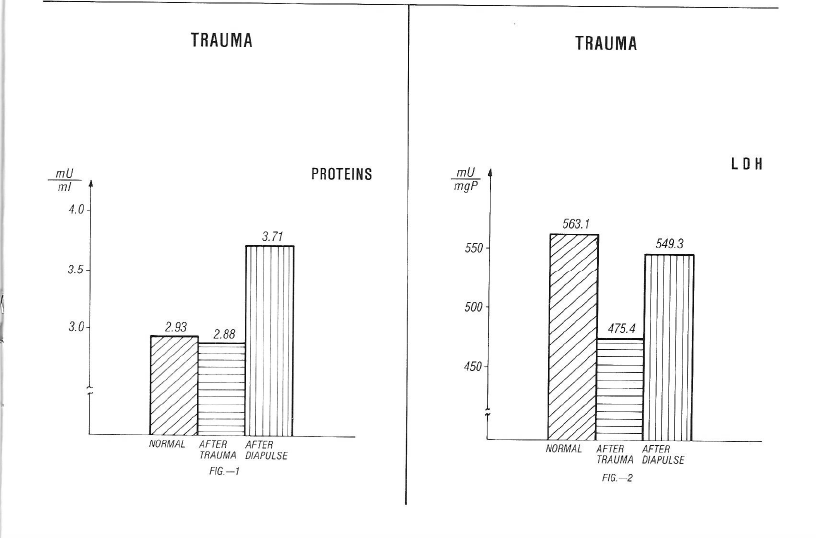
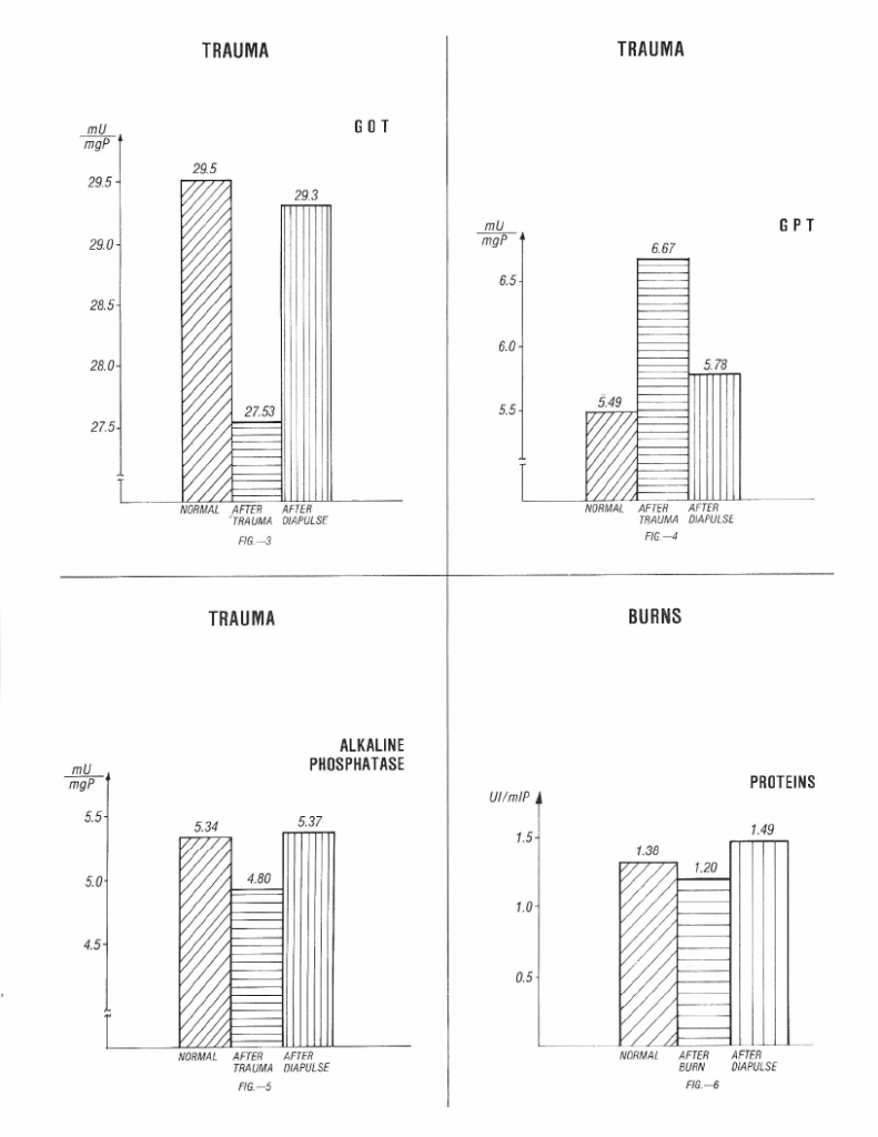
Fig 5 Alkaline Phosphatase / Fig 6 Proteins
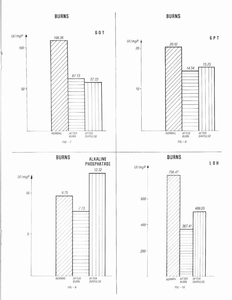
Fig 9 Alkaline Phosphatase / Fig 10 LDH